Radiofrequency Pulses
In order to create an image, the protons must be first knocked out of equilibrium. This is achieved by exciting the protons with an electromagnetic wave at the resonant frequency or the Larmor frequency. A typical hospital MRI has a B0 field strength of 1.5T, resulting in a Larmor frequency of 63.87 MHz. An electromagnetic wave with a frequency of MHz falls into the range of radiofrequency waves, and thus radiofrequency pulses are used to perturb protons away from the equilibrium state.
To visualize this effect, imagine trying to grab a child off a rapidly spinning merry-go-round in two situations.
1, You are standing in-place on the ground outside the merry-go-round and each time the child comes around the merry-go-round, you try to grab him.
2. When you see the baby pass, you start running around with the merry-go-round at the same pace, and then grab the baby while you are running.
Clearly, it is easier to save the child in the second scenario because you are moving at the same frequency as the merry-go-round and the child. With respect to MRI in this analogous situation, you are the radiofrequency pulse trying to push the proton (child) out of the merry-go-round (B0 field).
The degree to which the protons are tipped out of equilibrium is described by the flip angle. For the simplest example, we will assume that a 90 degree RF excitation is applied, though a wide variety of RF excitation angles can be used to tip the proton out of equilibrium a few degrees (12 - 15) or even invert the proton magnetization (180 degrees).
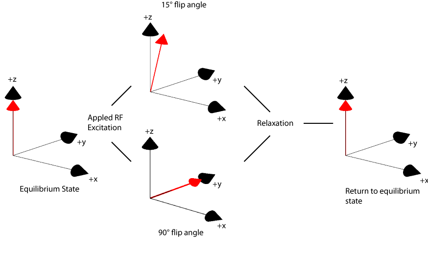
Figure: A radiofrequency pulse is applied in the yz-plane to tip the magnetization vector toward the +y axis. Since the vector is now out of equilibrium, it will start relaxing to return to its equilibrium state.
